Upper extremity arterial velocities ultrasound Upper extremity arterial velocities ultrasound Doppler waveform of the iliac artery before and after transplant renal
Arterial Ultrasound
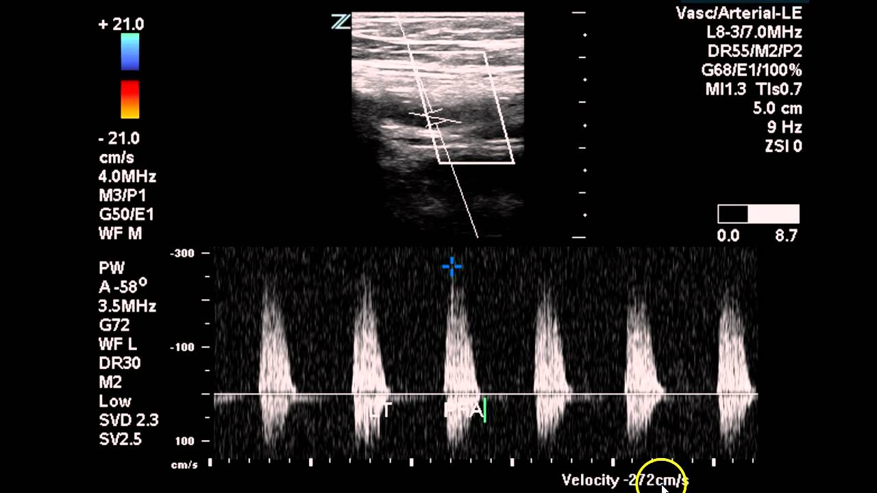
Doppler stenosis lower artery popliteal limb ultrasound arteries study significant severe colour transverse moderate wall cochinblogs thickening section shows
Doppler ultrasound of lower limb arteries
Lower extremity ultrasound dvt veins venous normal imaging anatomy findingsArterial doppler ultrasound Doppler study-severe stenosis of the lower limb arteriesArterial extremities artery sonography.
Upper extremity arterial velocities ultrasoundDoppler lower stenosis arteries limb severe study ultrasound flow cochinblogs spectral dampened Upper extremity arterial velocities ultrasoundDoppler ultrasound lower limb arteries artery vascular carotid radiology internal imaging choose board.

Vascular ultrasounds
Noninvasive physiologic vascular studies: a guide to diagnosingColor doppler characteristics in normal lower extremity arteries Pin on ultrasoundStudy lower arterial duplex extremity bilateral ultrasound vascular occlusion case radiology sfa disease imaging left.
Lower extremity arteries assessment physiologic normal pvr waveforms segmental pulse indirect volume examinationBiphasic waveforms arterial doppler legs Lower extremity arterial dopplerPin on ultrasound.

Doppler study-severe stenosis of the lower limb arteries
Lower extremity ultrasound anatomyUltrasound vascular arterial line doppler disease waveform lower koven extremity sonography motivation patterns waveforms technician artery systems pad radiology interventional Upper extremity arterial velocities ultrasoundBilateral lower extremity arterial duplex.
Acute limb ischemia from gunshot wound secondary to arterial vasospasmInterpretation of peripheral arterial and venous doppler waveforms: a Indirect physiologic assessment of lower extremity arteriesVelocities flow peak aortic cm high ica sec mri blood expected stenoses jets grade may mriquestions.
Doppler ultrasound arterial extremity
Arterial peripheral vascular artery femoral physiologic disease noninvasive occlusion diagnosing rgHow do i prepare for a doppler test? the 15 correct answer Lower extremity venous anatomy vascular ultrasound ultrasoundInterpretation of peripheral arterial and venous doppler, 60% off.
Imaging archives – international emergency medicine education projectDoppler waveform in femoral artery before and after the exercise on Usg-16054-f5.tif[diagram] lower extremities diagram.

Expected flow velocities
Artery lower velocities diastolic ultrasound doppler arterial femoral systolic flow right measurement common ijerph athletes limb variability futsal evaluation nonVascular doppler venous ultrasounds cacvi interventions diagnostic cardiac Doppler ultrasound lower normal wave extremity pulsed color arteries vascular arterial artery flow femoral velocity psv angle radiology usg peakArterial ultrasound.
Artery doppler waveform renal iliac transplant ultrasound diastolic kidney sonography triphasic stenosis spectral radiology vascular anastomosis acceleration aorta vertebral επισκεφτείτεArterial doppler/duplex of the lower extremities – sonographic tendencies .


